By the time a neurologist spots the classic resting tremor, the stooped posture, or the shuffling gait of Parkinson’s disease, the brain has already lost most of its ammunition. Researchers at Chalmers University of Technology put a hard number on it: 50 to 80 percent of the dopamine-producing neurons in the substantia nigra are already damaged or dead by the time major symptoms appear, according to the study’s first author, doctoral student Danish Anwer (News-Medical, Jan 2026). A 2025 Frontiers review puts the underlying degeneration on an even longer clock, noting that this neuronal loss unfolds over 5 to 15 years before diagnosis is even possible.
That lag is the central problem in Parkinson’s research today — and the reason a growing cluster of scientists across immunology, genomics, sleep medicine, and neurology are racing to find the earliest possible tell. What they’re discovering is that Parkinson’s doesn’t begin with a tremor. It begins quietly, in the gut, the immune system, the nose, and the sleeping brain — sometimes decades before a diagnosis is made.
The prodrome: a disease with a decades-long warning period
Neurologists call this silent stretch the “prodromal” phase — the period when biological damage is accumulating but hasn’t yet crossed the threshold into visible motor symptoms. According to the Movement Disorders Society’s prodromal criteria, this phase “commences several years before the onset of motor features,” and two groups of patients have become especially valuable for studying it: people with idiopathic REM sleep behavior disorder, and people who carry known Parkinson’s-linked genetic mutations but show no symptoms yet.
The prodromal window is not a minor scientific curiosity — it may be the single most important target for future treatment. As the Chalmers team put it, catching the disease in this window is “an important step towards facilitating early identification of the disease and counteracting its progression before it has gone this far” (News-Medical).
Clue 1: The disease may start in the gut, not the brain
One of the most influential — and still debated — theories in Parkinson’s research comes from German neuroanatomist Heiko Braak, who in 2003 proposed that alpha-synuclein, the misfolded protein at the heart of Parkinson’s pathology, doesn’t originate in the brain at all. Braak’s autopsy studies found Lewy body damage consistently in the vagus nerve and the enteric nervous system — the gut’s own neural network — sometimes ahead of damage in the midbrain (Parkinson’s Foundation).
The Braak hypothesis proposes that misfolded alpha-synuclein enters through the gut lining, aggregates in the enteric nervous system, and then travels backward up the vagus nerve into the brainstem before eventually reaching the substantia nigra — the region responsible for producing dopamine. This is consistent with the well-documented clinical observation that constipation and other gut symptoms often precede a Parkinson’s diagnosis by years.
Researchers at Johns Hopkins Medicine tested this directly by injecting misfolded alpha-synuclein into the stomachs of mice. As Ted Dawson, director of the Institute for Cell Engineering, explained, the pathologic protein “kind of rides up the vagus nerve into the brain,” after which “it spreads from cell to cell to create the symptoms of Parkinson’s disease” (Johns Hopkins Medicine). Supporting human evidence has followed: studies have linked truncal vagotomy (surgical cutting of the vagus nerve) and even prior appendix removal to a measurably lower risk of later developing Parkinson’s, since both procedures may interrupt the gut-to-brain highway the pathology seems to travel along.
Clue 2: The immune system may see it coming first
Perhaps the most striking recent finding comes from the La Jolla Institute for Immunology (LJI), where a team led by Professor Alessandro Sette has been tracking T cells — the immune system’s targeted attack cells — in people at high genetic risk of Parkinson’s. In a 2025 study published in npj Parkinson’s Disease, the researchers found that T cells reactive to alpha-synuclein and a second protein called PINK1 peak during the prodromal period, before diagnosis — not after (ScienceDaily).
“This T cell immunity could be a marker for early Parkinson’s treatment, even before people show symptoms,” Sette said, adding that “there’s reason to think that treating Parkinson’s in the very early stages can lead to a better outcome” (ScienceDaily).
The study tracked volunteers who already carried genetic risk factors and, in some cases, early prodromal signs such as disrupted REM sleep or loss of smell. Study co-author Emil Johansson framed the open question the team had been chasing: “We can see these reactive T cells in people after they develop Parkinson’s, but what happens before that?” (ScienceDaily). The answer — that T cell reactivity to PINK1 hits an all-time high right before diagnosis — raises a genuine chicken-and-egg question the field hasn’t resolved. Sette is careful not to overstate it: “Does that destruction cause autoimmunity — or is the autoimmunity the cause of the disease?” he asks. “That’s the chicken-and-the-egg of inflammation in Parkinson’s disease” (ScienceDaily).
Clue 3: A blood test that reads cellular stress, up to 20 years early
In January 2026, a joint team from Chalmers University of Technology and Oslo University Hospital published findings that may be the closest thing yet to a scalable early-warning blood test. Rather than looking for alpha-synuclein itself, they focused on two more upstream biological processes: DNA damage repair and cellular stress response — both of which appear to activate in a distinctive pattern during the prodromal window, and only during that window (News-Medical).
Using machine learning to sift through gene-activity data, the team identified a signature present in prodromal patients but absent in both healthy controls and patients with established, symptomatic disease. Study lead Annikka Polster, Assistant Professor at Chalmers, explained the significance: “we have found an important window of opportunity in which the disease can be detected before motor symptoms caused by nerve damage in the brain appear” (News-Medical). She added that the biomarkers “can be measured in blood,” which “paves the way for broad screening tests via blood samples: a cost-effective, easily accessible method” (News-Medical).
Critically, this window appears to close as the disease progresses — the same signature is no longer detectable once symptoms are established, which the researchers believe makes it not just a diagnostic clue but a potential map of the mechanisms driving early disease. The team believes blood tests built on this discovery could begin healthcare testing within roughly five years, though they caution that the gene activity measured in blood only partially mirrors what’s happening in the brain, and that the study population may not generalize to all patients (Fox News Health).
Clue 4: A 24-hour skin test for misfolded protein
While blood-based signatures look at the biological precursors of Parkinson’s, another line of research goes straight after the misfolded protein itself. The seed amplification assay (SAA) is a lab technique that can detect vanishingly small amounts of misfolded alpha-synuclein by using it as a “seed” to trigger a chain reaction of protein misfolding that becomes measurable over time.
Because alpha-synuclein pathology extends into peripheral nerves — including those in the skin — researchers have found they can run this assay on a simple skin biopsy rather than invasive cerebrospinal fluid extraction. A 2024 study published in npj Parkinson’s Disease optimized this skin-based approach and achieved a sensitivity of 92.46 percent and specificity of 93.33 percent across 332 Parkinson’s patients and 285 controls, with results available within 24 hours (Nature/npj Parkinson’s Disease). A subsequent 2025 systematic review found that across biological samples generally, skin-based SAA testing reached a pooled sensitivity of 0.91, trailing only extracellular vesicle-based assays and outperforming blood and cerebrospinal fluid on some measures (PMC meta-analysis).
The clinical appeal is obvious: unlike a lumbar puncture, a skin punch biopsy is quick, low-risk, and can plausibly be deployed at scale — including in people who have no motor symptoms yet but do have other prodromal red flags.
Clue 5: What happens in your sleep may predict what happens to your brain
Of all the prodromal markers identified so far, none has more consistent clinical backing than REM sleep behavior disorder (RBD) — a condition in which the muscle paralysis that normally accompanies dream sleep fails, causing people to physically act out their dreams, sometimes violently.
The Michael J. Fox Foundation notes plainly that RBD “can be one of the first symptoms of Parkinson’s, occurring years before a diagnosis,” alongside loss of sense of smell as another early warning sign. Research from the Foundation’s Parkinson’s Progression Markers Initiative (PPMI) has found that up to 41 percent of Parkinson’s patients experienced RBD before diagnosis (ABC News). Longitudinal studies of people with isolated RBD — meaning no other neurological diagnosis yet — show that roughly 80 to 90 percent eventually convert to a synucleinopathy such as Parkinson’s or dementia with Lewy bodies (medRxiv GWAS study).
Dr. Rachel Dolhun, a movement disorder specialist and head of medical communications at the Michael J. Fox Foundation, has emphasized why this population matters so much to researchers: “People who live with RBD can help researchers understand how and why Parkinson’s comes on from the very earliest moments” (ABC News). One recent biomarker study of people with confirmed RBD found that a combination of three prodromal signals — smell loss (hyposmia), alpha-synuclein SAA positivity, and abnormal dopamine transporter (DAT) imaging — together helped predict which RBD patients were most likely to “phenoconvert,” or develop clinical Parkinson’s, in the near term (PMC, 2025).
Clue 6: Imaging the dopamine system before it fails
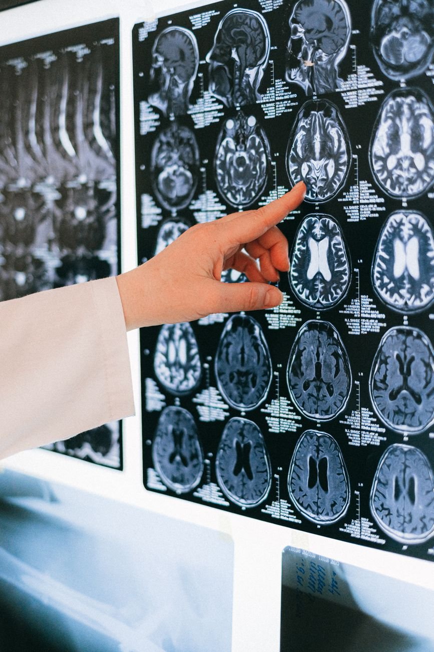
Brain imaging remains one of the more established prodromal tools, particularly DAT-SPECT scans, which visualize dopamine transporter density in the striatum. Reduced striatal DAT binding is strongly associated with eventual conversion to Parkinson’s in at-risk populations, and researchers studying RBD patients have found DAT positivity in roughly 80 percent of those who went on to develop the disease, compared with only about 10 percent of those who didn’t (PMC, 2025). Circuit-level imaging research more broadly is trying to characterize how basal ganglia and cortical networks shift during the preclinical and prodromal stages, well before a movement disorder specialist would flag anything abnormal on a standard exam (PMC review, circuit imaging biomarkers).
Why none of this is a single “Parkinson’s test” — yet
It’s worth being direct about where the science actually stands. No single biomarker discussed here — not the T cell signature, not the blood-based stress-response pattern, not the skin SAA, not RBD, not DAT imaging — is currently used on its own to diagnose Parkinson’s in clinical practice. Diagnosis today still relies primarily on clinical history and physical examination (ABC News). Each of these markers also comes with real limitations: the Chalmers group notes their blood signal only partially reflects brain biology; the LJI team is careful to say their T cell findings don’t prove causation; and skin SAA struggles to distinguish disease subtypes even when it reliably flags disease presence (European Journal of Neurology, 2025).
What’s changed is the direction of travel. Researchers are increasingly combining these signals — genetic risk, smell loss, RBD, imaging, alpha-synuclein assays, and now immune and blood-based markers — into composite risk models, such as the MDS Prodromal PD Probability score, rather than hunting for one silver-bullet test. Sette’s own lab is already extending this framework to Alzheimer’s, noting that “a lot of progress has been made toward identifying people in very early stages of the disease progression” in that field too (ScienceDaily) — a sign that the same prodromal-detection playbook may soon apply across neurodegenerative disease broadly.
The stakes of catching it early
With more than 10 million people living with Parkinson’s worldwide, and that number expected to more than double by 2050 as populations age (News-Medical), the push toward prodromal detection isn’t just an academic exercise. Every biomarker discussed here is ultimately being chased for the same reason: today’s Parkinson’s treatments manage symptoms, but by the time they start, most of the relevant brain tissue is already gone. Catching the disease in its silent decades — while there are still dopamine neurons left to protect — is what researchers across immunology, sleep medicine, and neurogenomics increasingly see as the real opportunity for turning Parkinson’s from a disease we manage into one we might eventually prevent.


No responses yet